Host immune surveillance plays a crucial role in recognizing and destroying invading pathogens and host cells that become cancerous, so immunotherapy has become one of the major methods for cancer prevention and treatment. Accumulating evidence implicates that Ganoderma lucidum polysaccharides(GLP) exert the anticancer action in part by stimulating the immune function.
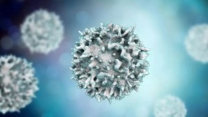
GLP can activate T and B lymphocytes, macrophages, dendritic cells (DCs), and natural killer (NK) cells, which promote lymphocyte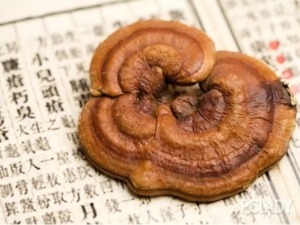 proliferation, enhance phagocytosis, and increase cytokine production augments NK cell-mediated cytotoxicity.
-
Effect of GLP on T lymphocytes
T lymphocytes (also known as T cells) are essential for cellular immunity. There are several subtypes of T cells with different functions, such as T helper cells, cytotoxic T cells, memory T cells, suppressor T cells, natural killer T cells, and mucosal-associated invariant T cells.
Numerous studies have suggested that GLP is an activator of T lymphocytes. The treatment with GLP significantly promotes concanavalin A (ConA)-induced mouse lymphocyte proliferation and IL-2 production.
GLP can also enhance the DNA synthesis in mouse spleen cells in a mixed lymphocyte culture by inducing the expression of DNA polymerase α. GLP increases the expression of IFN-γ in the T-lymphocytes and IL-1, IL-2, and IFN-γ in mouse spleen cells. GLP increases the production of inositol triphosphate (IP3) and diacylglycerol (DAG) in resting T cells.
However, it does not influence the production of IP3 and DAG in ConA-activated T cells, suggesting that both IP3/Ca2+ and DAG/ protein kinase C (PKC) pathways may be involved in the immunomodulatory effect of GLP on T cells. More studies have exhibited that GLP can activate PKC and protein kinase A (PKA) in murine T cells.
B16F10 melanoma cells can secrete a large amount of interleukin 10 (IL-10), transforming growth factor β1 (TGF-β1) and vascular endothelial growth factor (VEGF) in the cell culture medium. The treatment with the B16F10 cell culture supernatant prevents phytohemagglutinin (PHA) from stimulating the production of perforin and granzyme B and the proliferation in lymphocytes.
GLP can fully or partially antagonize the inhibitory effects of the B16F10 cell culture supernatant on lymphocytes. GLP also prevents B16F10 cells from reducing the expression of CD71 and Fas ligand (FasL) in lymphocytes.
Furthermore, the plasma of lung cancer patients inhibits proliferation, CD69 expression, and perforin and granzyme B production in lymphocytes activated by PHA, which can also be fully or partially reversed by GLP treatment.
 GLP treatment of the co-culture of B16F10 melanoma cells and lymphocytes increases the production of CD69 (a co-stimulatory molecule for T cell activation and proliferation), FasL, and interferon (IFN)-γ in the medium.
-
Effect of GLP on B lymphocytes
B lymphocytes (also known as B cells) play an important role in humoral immunity. Unlike T or NK cells, B cells express B cell receptors on their cell membrane, allowing the B cell to bind a specific antigen and produce an antibody against the antigen.
B cells also present antigens and secrete cytokines. GLP can activate B cells by increasing their proliferation and differentiation. GLP increases the percentage of B cells by 2.5–4 fold and increases the size of B cells.
In vivo treatment with GLP also activates spleen and bone marrow-derived B lymphocytes from sarcoma S180 bearing mice and induces proliferation of the B cells and production of large amounts of immunoglobulins in mice. GLP can induce the expression CD71 and CD25 on B cell surface and increase the secretion of immunoglobulins by B cells by directly stimulating the expression of PKCα and PKCγ in B cells.
-
Effect of GLP on DCs
DCs are important professional antigen-presenting cells that are necessary to initiate the primary immune response of both helper and cytotoxic T lymphocytes. GLP can stimulate the maturation of normal human monocyte-derived DCs and leukemic monocyte-derived DCs. GLP increases cell-surface expression of CD80, CD86, CD83, CD40, CD54, and human leukocyte antigen-DR (HLA-DR).
It also enhances the production of IL-12 p70, IL-12 p40, and IL-10. The GLP-induced activation and maturation of human monocyte-derived DCs are mediated by the nuclear factor kappa-light-chain-enhancer of activated B cells (NF-κB) and p38 mitogen-activated protein kinase (MAPK) pathways.
GLP (0.8, 3.2, or 12.8 mg/ml) increases the co-expression of CD11c, and I-A/I-E molecules on the surface of cultured bone marrow-derived DCs. It elevates mRNA expression of cytokine IL-12 p40 in DCs and increases protein production of IL-12 p40 in culture supernatants.
GLP also enhances the lymphocyte proliferation of mixed lymphocyte cultures induced by mature DCs. Treatment of leukemic monocytic cell lines THP-1 and U937 with GLP (100 μg/mL) significantly increases the expression of HLA-DR, CD40, CD80, and CD86. GLP induces THP-1 leukemia cells to macrophage-like cells by enhancing cell adherence, superoxide production, cell cycle arrest, and expression of differentiation markers such as CD11b, CD14, CD68, and matrix metalloproteinase-9 (MMP-9), and myeloperoxidase. Also, GLP-induced activation of caspases and p53 contributes to this differentiation.
-
Effect of GLP on macrophages
Macrophages are the “big eaters” of the immune system, which engulf and digest apoptotic cells and pathogens (called phagocytosis), and produce immune effector molecules.
In vivo treatment with GLP activates bone marrow-derived macrophages from sarcoma S180-bearing mice, resulting in the production of immunomodulatory substances, such as IL-1β, TNF-α, and nitric oxide (NO). GLP increases phagocytosis of macrophages significantly and enhances macrophage-mediated tumor cytotoxicity.
Also, GLP activates macrophages in vitro and increases the levels of various cytokines, including IL-1β, tumor necrosis factor (TNF)-α, IFN-γ, and IL-6 in the culture medium. GLP has been shown as an inducer of MAPKs- and Syk-dependent TNF-α and IL-6 secretion in murine resident peritoneal macrophages. GLP stimulates Dectin-1, but toll-like receptor (TLR)-4 signaling is not involved in the biological activities of GLP.
However, this is controversial since GLP has been found to directly bind to TLR-4, mIg of B cells, 7S ribosomal protein, and bZIP enhancer, and to transduce certain signalings via the TLR-4. GLP also induces the expression of inflammatory cytokine IL-1, which partially links to its anticancer activity. GLP up-regulates the secretion of IL-1 and the expression of pro-IL-1 (precursor of IL-1) and IL-1-converting enzyme in human macrophages and murine macrophages (J774A.1). This is attributed to the activation of protein tyrosine kinase/protein kinase C/MEK1/extracellular signal-regulated kinase (ERK) and protein tyrosine kinase/Rac1/p21-activated kinase/p38 pathways.
-
Effect of GLP on NK cells
NK cells, unlike cytotoxic T-cells, can recognize stressed cells in the absence of antibodies and major histocompatibility complex (MHC), allowing for a much faster immune reaction.
Hence, NK cells are critical to innate immunity. It has been shown that GLP increases the population of CD14+CD26+ monocyte/macrophage, CD83+CD1a+ DCs, and CD16+CD56+ NK cells by 2.9, 2.3, and 1.5 fold, respectively, in human umbilical cord blood mononuclear cells.
Also, GLP enhances NK cell-mediated cytotoxicity by 31.7%. Oral administration of G. lucidum extract increases T-helper type 1 and macrophage cytokines (IL-6 and IFN-γ) and enhances the NK cell activities and phagocytosis in BALB/c mice.
A GLP fraction binds to the TLR-4 receptor and activates ERK, c-Jun N-terminal kinase (JNK), and p38 MAPK. GLP can stimulate the expression of IL-1, IL-6, IL-12, IFN-γ, TNF-α, granulocyte-macrophage colony-stimulating factor (GM-CSF), granulocyte-colony stimulating factor (G-CSF), and macrophage colony-stimulating factor (M-CSF) in mouse splenocytes.
Additionally, a GLP fraction increases the number of DCs and CD4, CD8, regulatory T, B, plasma, NK, and natural killer T (NKT) cells in the spleen of mice.
In vivo treatment of mice with this fraction elevates the levels of 12 cytokines and chemokines, including KC (CXCL1), MCP-1 (CCL2), IL-6, MIP-1β (CCL3), IL-1 β, IL-12p40, IL-12p70, RANTES (CCL5), IL-1α, TNF-α, IL-10, and IL-13 in the serum of mice.
Ganoderma lucidum mitigates cyclophosphamide-induced decrease in body weight, NK activity, IFN-γ production, and cytotoxic T lymphocyte activity and inhibits the abnormal increase or decrease in IL-4 level due to cyclophosphamide administration.
Chronic administration of 2.5 mg/kg GLP accelerates recovery of bone marrow cells, red blood cells, white blood cells, and splenic NK cells and NKT cells. It enhances T and B cell proliferation responses compared to the treatment with vehicle.
It also increases the phagocytosis and cytotoxicity of macrophages without any apparent side effects. Thus, the low-dose GLP treatment accelerates the recovery of immunosuppressed mice from leukopenia, myelosuppression, and immunosuppression, which should be beneficial to cancer chemotherapy.
Leave A Comment