In recent years, traditional herbal medicines have gained more interest as a complementary and alternative approach to cancer therapy because of their insignificant side effects on the human body. Ganoderma lucidum (GL), an important natural product derived from the medicinal mushroom, draws attention since it enhances human health and increases longevity. It has exhibited significant antitumor effects against different types of tumors via multiple mechanisms involving anti-proliferation, pro-apoptosis, anti-metastasis, and anti-angiogenesis. We evaluated GL’s cancer preventive and anti-immunosuppression properties in the present study using established skin carcinogenesis models.
The non-tumorous murine epidermal JB6 P+ cells and UV-induced murine skin carcinogenesis are well-established models for investigating the mechanism of skin carcinogenesis and identifying natural or synthetic chemopreventive agents. We first used the in vitro assay of JB6 P+ transformation induced by the tumor promoter EGF to examine the effects of gastrointestinal extracts of GLSF and its component GLS.
Since the data showed a highly potent anti-transformation activity for both extracts, in vivo studies were also conducted in SKH-1 mice. The tumors in UV-irradiated SKH-1 mice imitate human skin SCC formation and reflect clearly defined tumor formation stages. Dietary GLSF treatment decreased the tumor incidence and reduced the average number of tumors per mouse, indicating a protective activity against carcinogenesis.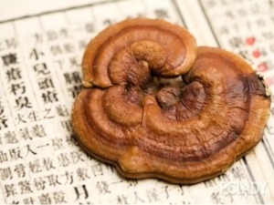
Furthermore, epidermal thickness and histopathological observations of the non-tumor skin indicate that GLSF supplementation reversed the chronic inflammation caused by UV and showed nearly habitual architecture, which might contribute to the decrease in tumor formation, implying the antitumor action of GLSF. However, GLS and GLF did not show the same degree of prevention as GLSF, indicating that the combination of these two components may synergistically contribute to the activity of GLSF.
The Ki-67 is a marker usually used to estimate cellular proliferation in different cancers, including skin cancer. It is a protein that consists of two isoforms having molecular weights of 345 and 395 kDa, and its expression level persists high through all active phases of the cell cycle, including G1, S, G2, and M, but low in the resting phase (G0).
Therefore it is considered a clinically vital index to show the proliferation capacity of tissues. The expression of Ki-67 is also correlated with the proliferative activity of tumor cell populations and is commonly used as a marker for tumor aggressiveness. A reduction in the expression of this marker is linked with a response to cancer treatment. Numerous cancer studies have examined the prognostic importance of Ki-67 IHC staining, including in skin cancer. In our research, GLSF significantly ameliorated Ki-67-positive cells in the non-tumor and tumor skin tissue compared with the UV-only group. Our study is consistent with the previous report in which GL treatment decrease Ki-67 cell expression.
In our study, we found that GLSF treatment decreased the expression of COX-2 and NF-kB significantly as compared to the only UV group. Our results indicate that inflammation is a major target for GLSF and previous studies validate our data. Inflammation is the body’s response to tissue damage and coordinates with cellular transformation and the immune system to help repair injured tissue. Different types of mediators produced by immune cells during the inflammation play an important role in making homeostasis balance.
However, in chronic inflammation, overproduction of immune mediators can disrupt this homeostasis balance and secrete excessive pro-inflammatory agents, including prostaglandin E2 (PGE2), via up-regulating the Cox-2 activity, which leads to inflammation-mediated diseases, including cancer. Cox-2 and its product PGE2 play an important role in the skin’s response to UV radiation which Cox-2 knockout mice have demonstrated. Cox-2 inhibitors such as nonsteroidal anti-inflammatory drugs (NSAIDs) have shown chemopreventive activity against UV-induced skin carcinogenesis.
Thus, our data indicate that GLSF prevents skin cancer via targeting COX-2 expression. Although the exact mechanism is unknown, since GL is a non-toxic dietary supplement, it offers improved safety advantages compared with nonsteroidal anti-inflammatory drugs.
Activation of Nuclear factor-κB (NF-κB) has been linked to various cellular processes in cancer, including inflammation and proliferation. It is a transcription factor that governs many genes associated with multiple immune and inflammatory responses, including skin inflammation. The inactive NF-κB exists as a heterodimer of the p65 and p50 subunits in the cytoplasm, which are bound to the inhibitory protein IκB. Activation of NF-κB can be induced directly by UV or pro-inflammatory cytokines such as tumor necrosis factor and IL-1.
Therefore, drugs that inhibit NF-κB activity have been helpful as a chemopreventive agent in various cancers. The p65 subunit of NF-κB is essential for skin carcinogenesis in mice since the loss of p65 in the keratinocytes prevented both SCC tumor initiation and tumor promotion. GLSF treatment significantly attenuated NF-κB expression in UV-exposed mice, suggesting it has potent anti-inflammatory activity.
UV-induced immunosuppression has been linked to the development of photocarcinogenesis. We investigated the efficacy of GSLF on UV-induced immunosuppression using a local contact hypersensitivity assay in mice. Contact hypersensitivity (CHS) is an immune cell-dependent T cell-mediated response elicited by various sensitization chemicals. A hypersensitivity in mice and humans is activated by Th1/T cytotoxic-1 effector cells and downregulated by Th2/T regulatory (Treg) CD4+ T cells. The role of GLSF in UVB-induced immune suppression of CHS is unknown.
These mechanisms are of significant clinical importance as UV-induced immunosuppression has been associated as a risk factor for nonmelanoma skin cancers. To test the effect of GLSF in UV-induced immunosuppression, we treated GLSF to mice one month before UV exposure. The results showed that GLSF reversed the immunosuppression effect of UV effectively. Further studies are needed to identify the active components in GLSF responsible for such cancer preventive and immunomodulatory activity.
In conclusion, our data strongly prove that dietary supplements of GLSF may have preventive effects against UV-induced skin tumor development, which may be partly attributed to GLSF’s activity in the modulation of cutaneous immunity.
Leave A Comment