- Anti-Oxidation
Carotenoids contain long conjugated double bonds in a polyene chain responsible for antioxidant activities by quenching singlet oxygen and scavenging radicals to terminate the chain reaction. Astaxanthin has a conjugated polyene chain at the center and hydroxy and keto moieties on each ionone ring. Due to its unique molecular structure, astaxanthin shows better biological activity than other antioxidants because it can link with the cell membrane from the inside to the outside. The polyene chain in astaxanthin traps radicals in the cell membrane. In contrast, the terminal ring of astaxanthin can scavenge radicals both at the surface and in the interior of the cell membrane.
Astaxanthin is more effective than β-carotene in preventing lipid peroxidation in solution and biomembrane models such as egg yolk phosphatidylcholine liposomes and rat liver microsomes. Astaxanthin was approximately twofold more effective than β-carotene in inhibiting liposome peroxidation induced by ADP and Fe2+. Astaxanthin could trap radicals at the conjugated polyene chain and in the terminal ring moiety.
The proposed molecular interaction was as follows: the two-terminal rings interact with the hydrophilic polar site of membrane phospholipids, and the hydroxyl and carbonyl groups form an intramolecular hydrogen-bonded five-membered ring, increasing the hydrophobicity of astaxanthin. It is well known that the activity of carotenoids can be shifted from antioxidant to pro-oxidant according to their concentrations, high oxygen tension, or interactions with other co-antioxidants. 17 carotenoids were divided into three classes:
(1) those without significant antioxidative properties;
(2) those with good antioxidative but also pro-oxidative properties; and
(3) those with antioxidative solid and without any pro-oxidative properties. Astaxanthin was categorized as class (3), whereas β-carotene and lycopene were identified as class (2).
The increase in the susceptibility of low-density lipoprotein (LDL) and cell membrane lipids to oxidative processes contributes to atherosclerosis and thrombus formation. Our group was the first to report that astaxanthin protected human LDL against oxidative attack. Compared to α-tocopherol and lutein, astaxanthin showed a greater antioxidative effect on LDL oxidation induced by AMVN-CH3O (2,2-azobis-4-methoxy-2,4-dimethylvaleronitrile) in vitro. To confirm the antioxidant effect of astaxanthin ex vivo, we recruited 24 healthy adults to consume astaxanthin purified from krill at 0, 1.8, 3.6, 14.4, and 21.6 mg/day for 14 days and then measured the changes in LDL oxidizability. At the end of the study, astaxanthin consumption significantly prolonged the lag time, a marker of the susceptibility of LDL to oxidation, at the dose levels of 3.6 mg/day and higher.
Importantly, an intake of 3.6 mg astaxanthin is equivalent to approximately 165 g of salmon flesh. Researchers reported the efficacy of astaxanthin supplementation (6 and 12 mg/day) on phospholipid hydroperoxides (PLOOH) levels in erythrocytes in 30 healthy subjects. After 12 weeks of administration, decreased PLOOH levels and increased astaxanthin in erythrocytes were observed.
Researchers also reported that astaxanthin supplementation for 12 weeks reduced the revels of plasma 12- and 15-hydroxy fatty acids in healthy males. In obese and overweight adults, supplemental astaxanthin (5 and 20 mg/day) reduced biomarkers of oxidative stress, including malondialdehyde (MDA) and isoprostane, and increased superoxide dismutase (SOD) and total antioxidant capacity (TAC). These findings suggest that astaxanthin may decrease in vivo lipid peroxidation.
Several antioxidant enzymes catalyze reactions to counteract free radicals and ROS. Nuclear factor erythroid-related factor 2 (Nrf2) is a master regulator of the antioxidant response and xenobiotic metabolism through regulating a wide range of antioxidant and Phase II detoxification genes. Astaxanthin treatment attenuated cyclophosphamide-induced oxidative stress, DNA damage, and cell death in rat hepatocytes through an Nrf2-antioxidant response element (ARE) pathway.
It was further observed that the levels of Nrf2 and the targeted phase-II enzymes, i.e., NAD(P)H dehydrogenase, quinone 1 (NQO1), and heme oxygenase 1 (HO-1), were increased with astaxanthin treatment. Interestingly, astaxanthin showed synergistic effects on the induction of the cellular glutathione level and the mRNA expression of Nrf2 and its target genes (NQO1, HO-1, and glutathione S-transferase mu 2 [GSTM2]) when combined with docosahexaenoic acid (DHA) or eicosapentaenoic acid (EPA) in a human hepatoma cell line.
Paraoxonase 1 (PON1), which is mainly responsible for the breakdown of lipid peroxides before they can accumulate in LDL, is an enzyme located on circulating high-density lipoprotein (HDL) particles. Still, oxidants readily inactivate the enzymatic activity of PON1. In hypercholesterolemic rabbits, astaxanthin prevented protein oxidation and changes in PON1 and thioredoxin reductase (TrxR-1) activities. TrxR-1 is a redox-active protein that efficiently regenerates oxidized thioredoxin.
These effects were not related to a direct effect of astaxanthin on these enzymes because astaxanthin enhanced TrxR-1 and had no effect on PON1 activity in vitro. It was reported that regular physical activity might increase PON1 activity. Astaxanthin supplementation (4 mg/day) for 90 days showed a beneficial effect in improving PON1 activity as well as the total sulphydryl group content in young soccer players. The same researchers also reported that post-exercise creatine kinase (CK) and aspartate aminotransferase (AST) levels were significantly lower in their astaxanthin group compared to a placebo group. Astaxanthin might be of special interest to athletes more susceptible to oxidative stress.
- Anti-Inflammation
Chronic inflammation is the main pathophysiological factor in many diseases, such as diabetes, hypertension, and atherosclerosis. Inhibiting the production of intracellular ROS is a general way to suppress the proinflammatory signals, and thus modulators of redox balance are considered the key regulators of inflammatory responses. Macrophages play a central role in inflammation and atherosclerosis progression. The scavenger receptor-mediated uptake of oxidized-LDL by macrophages leads to the formation of foam cells and the development of atherosclerotic plaque. The class A scavenger receptor (SR-A) and CD36 are responsible for the major part of oxidized LDL uptake by macrophages, suggesting the pro-atherogenic roles of SR-A and CD36.
Additionally, in inflammation states, macrophages produce excess amounts of matrix-degrading enzymes, proinflammatory cytokines/chemokines, nitric oxide (NO), and cyclooxygenase-2 (COX-2). Matrix metalloproteinases (MMPs), a family of Zn2+-dependent endopeptidases, are responsible for the degradation of most extracellular matrix proteins, and they mediate tissue remodeling in various pathologic conditions. The expression of MMPs is increased in atherosclerotic lesions and is linked to the weakening of the vascular wall due to degradation of the extracellular matrix.
In our previous study, astaxanthin remarkably suppressed the expression of the scavenger receptors SR-A and CD36), the activity and the expression of MMPs, and the mRNA expression of various inflammatory mediators, i.e., tumor necrosis factor (TNF)-α, interleukin (IL)-1β, IL-6, inducible nitric oxide synthase (iNOS), and COX-2, in THP-1 macrophages. We speculated that the antioxidant property of astaxanthin might account for its inhibitory effects on macrophage inflammation through the inhibition of nuclear factor-kappa B (NF-κB) activation. The proinflammatory cytokines, prostaglandins, and NO produced by activated macrophages play critical roles in atherogenesis.
The inhibition of proinflammatory cytokine secretion from macrophages might be one of the mechanisms mediating the beneficial effects of antioxidants on atherosclerosis development. Another study reported that astaxanthin decreased the expression of proinflammatory mediators such as prostaglandin E2, TNF-α, and IL-1β by suppressing IκB-dependent NF-κB activation in both lipopolysaccharides (LPS)-stimulated RAW264.7 cells and primary macrophages.
Astaxanthin significantly reduced the production of proinflammatory cytokines (TNF-α and IL-6) in LPS-stimulated neutrophils. Their study also revealed that astaxanthin enhanced neutrophil phagocytic and microbicidal capacity and suppressed superoxide anion and hydrogen peroxide production, which might be mediated by calcium released from intracellular storage and NO production.
These studies indicated that the anti-inflammatory effects of astaxanthin through its suppression of NF-κB activation may be based on its antioxidant activity. In addition, astaxanthin treatment reduced the secretion of proinflammatory cytokines (IL-1β, IL-6, and TNF-α) in H2O2-stimulated U937 mononuclear cells. This property was elicited by restoring the basal SHP-1 protein expression level and reducing NF-κB (p65) nuclear expression. SHP-1 is a protein tyrosine phosphatase that acts as a negative regulator of inflammatory cytokine signaling, and SHP-1 deficiency in mice causes spontaneous inflammation and autoimmunity; the authors of that study proposed that astaxanthin most likely inhibits the ROS-induced production of NF-κB transcription factor through the restoration of physiological levels of SHP-1.
Ischemia-reperfusion (IR) is a complex inflammatory process that includes major oxidative stress induced by ischemia and hypoxia. Astaxanthin treatment significantly decreased the conversion of xanthine dehydrogenase (XDH) to xanthine oxidase (XO), which reduced the level of oxidative stress in hepatocellular injury following IR in rats.
Another study showed that apoptosis and autophagy caused by hepatic IR injury were attenuated by astaxanthin pretreatment following a reduction in the release of ROS and inflammatory cytokines via the mitogen-activated protein kinase (MAPK) pathway in mice. Some studies reported astaxanthin’s protective effects on the brain and myocardial injury following IR.
In a clinical study, we studied the possible immune-enhancing, antioxidative, and anti-inflammatory activity of astaxanthin in healthy adult females, and their results showed that astaxanthin supplementation (2 and 8 mg/day) for eight weeks could decrease both the level of DNA oxidative damage biomarker and inflammation, and enhance the immune response. The enhancement of the immune response was also observed in dogs fed astaxanthin, β-carotene, and lutein.
- Lipid Metabolism
Astaxanthin has been reported to improve dyslipidemia and metabolic syndrome in animal models. In apoE knockout mice fed a high-fat and high-cholesterol diet, astaxanthin increased the levels of LDL receptor (LDLR), 3-hydroxy-3-methylglutaryl CoA (HMG-CoA) reductase, and sterol regulatory element-binding protein 2 (SREBP-2) in the liver, which might be responsible for the hypocholesterolemic effect of astaxanthin. In the same experiment, the expressions of carnitine palmitoyl transferase 1 (CPT1), acetyl-CoA carboxylase β (ACACB), and acyl-CoA oxidase (ACOX) mRNA were significantly increased by astaxanthin supplementation, suggesting that the triglyceride-lowering effect of astaxanthin might be due to increased fatty acid β-oxidation in the liver.
We investigated the effects of astaxanthin on key molecules in cholesterol efflux from macrophages. The study revealed that astaxanthin did not modify the peroxisome proliferator-activated receptor (PPAR)-γ or liver X receptor (LXR)-α and -β levels. Still, it increased the expression of ATP-binding cassette transporters (ABC) A1 and G1, thereby enhancing the cholesterol efflux from macrophages.
In diet-induced obesity in mice, astaxanthin significantly lowered the plasma triglyceride, alanine transaminase (ALT), and AST levels. It increased the mRNA expression of antioxidant genes regulated by Nrf2 in the liver. In addition, astaxanthin decreased macrophage infiltration and apoptosis of vascular cells in atherosclerotic plaques and provided stabilization of the plaques in hyperlipidemic rabbits.
To determine the lipid metabolism-modulating effect of astaxanthin in humans, we conducted a placebo-controlled study of astaxanthin administration at doses of 0, 6, 12, and 18 mg/day for 12 weeks with 61 non-obese subjects with mild hyperglycemia. Multiple comparison tests showed that 12 and 18 mg/day of astaxanthin significantly reduced the subjects’ triglyceride levels, and the 6- and 12-mg doses significantly increased HDL cholesterol.
The serum adiponectin level was also increased by astaxanthin (12 or 18 mg/day), and the changes in adiponectin positively correlated with the HDL-cholesterol changes. The HDL-increasing effect of astaxanthin is thus of significant interest because a very limited number of dietary factors were suggested to increase HDL-cholesterol concentrations. Our study showed a markedly positive correlation between the percentage change of adiponectin and the HDL-cholesterol level.
Although the mechanisms of astaxanthin-mediated adiponectin elevation are still poorly understood, several investigations have shown a significant and independent association between serum adiponectin and HDL cholesterol and that adiponectin may directly regulate HDL metabolism through a dual effect on the very-low-density lipoprotein (VLDL)-triglyceride pool and hepatic lipase.
Another mechanism of the astaxanthin-induced increase in HDL-cholesterol may be considered, albeit by an implicit action, due to the increased ABCA1 expression and cholesterol efflux from macrophages through the actions of adiponectin increased by astaxanthin.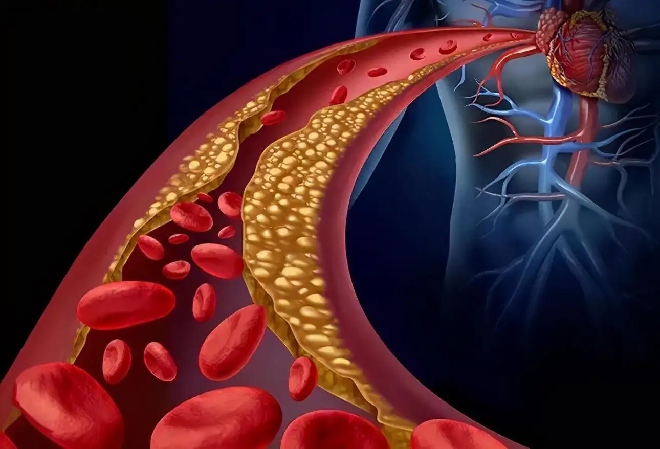
In contrast, a recent meta-analysis of seven randomized controlled trials failed to identify a significant effect of astaxanthin on the plasma lipid profile, but a slight glucose-lowering effect was observed. The review’s authors mentioned that the study interpretation had limitations regarding the heterogeneous populations, the varying concepts of the studies, and the different quantities of astaxanthin used.
Several studies have indicated that changes in the intracellular redox balance can modify lipid metabolism. Indeed, oxidative stress was associated with lipid accumulation in adipose tissue and affected the regulation of hepatic lipid synthesis. During exercise, ROS generation in skeletal muscle increases along with the elevation of energy expenditure. ROS may also affect the utilization of energy substrates in muscle, leading to a disorder in lipid metabolism.
Astaxanthin accelerated lipid utilization during exercise, leading to improved endurance and efficient body fat reduction with training in mice. An increase of fatty acyl-CoA uptake into the mitochondria via CPT1 during exercise may be involved in the promotion of lipid metabolism by the antioxidant activity of astaxanthin. According to these findings, astaxanthin is expected to improve aerobic performance and body weight control by modifying muscle energy metabolism via its antioxidant effect.
- Glucose Metabolism and Blood Pressure Control
Growing evidence suggests that diabetes and other disorders of glucose metabolism, such as impaired glucose tolerance (IGT), need to be considered independent risk factors for CVD. Since oxidative stress promotes insulin resistance in obesity and type 2 diabetes, it is crucial to find effective antioxidants to decrease this threat.
As mentioned above, a recent meta-analysis of randomized controlled trials showed that astaxanthin supplementation slightly lowered glucose levels. The antidiabetic effects of astaxanthin could be explained by means of several proposed mechanisms. In db/db mice, astaxanthin showed a protective effect against oxidative stress and cytotoxicity in pancreatic β-cells. Astaxanthin activated the hepatic IRS-PI3K-Akt signaling pathway and improved glucose metabolism in the liver of high-fructose and high-fat diet (HFFD)-fed mice.
Another study reported that astaxanthin treatment normalized the activities of hexokinase, pyruvate kinase, glucose-6-phosphatase, fructose-1,6-bisphosphatase, and glycogen phosphorylase and increased the glycogen reserves in the liver of HFFD-fed mice. In addition, astaxanthin decreased the HFFD-induced activation of serine kinases (JNK and ERK).
The anti-obesity effect of astaxanthin has been reported in high fat-fed mice; astaxanthin was shown to increase fatty acid utilization, which can be responsible for its anti-diabetic effect. It is known that fucoxanthin, a xanthophyll carotenoid present in brown seaweeds, induces uncoupling protein 1 (UCP1) in mitochondria, leading to the oxidation of fatty acids and heat production in white adipose tissue.
Studies in animal models of insulin resistance and fatty liver have demonstrated that hepatic steatosis and endoplasmic reticulum (ER) stress are linked. A recent study showed that disruption of ER homeostasis led to chronic unfolded protein response (UPR) and induced inflammation and insulin resistance in the liver. Hepatic ER stress can promote de novo lipogenesis, while lipids can exacerbate ER stress, a situation that creates a vicious cycle. Astaxanthin reduced hepatic ER stress, ROS production, phosphorylation of JNK, and NF-κB-mediated inflammation in HFFD-fed mice.
Astaxanthin may also have prevented the progression of diabetic nephropathy by decreasing renal oxidative stress and preventing renal cell damage in db/db mice. Another study reported that astaxanthin beneficially affected sucrose-induced blood pressure elevations and insulin resistance at relatively high doses in rats. Astaxanthin may have an innate antihypertensive effect because astaxanthin administration lowered the blood pressure and delayed the incidence of stroke in spontaneously hypertensive rats (SHRs).
Leave A Comment