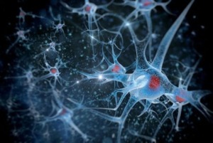
The human Central Nervous System (CNS) contains 100 billion neuronal cells and an equal number of glia cells, such as microglia, astrocytes, and oligodendrocytes. The CNS includes all nerves in the brain and spinal cord and is isolated from the other compartments of the human body through the blood-brain barrier (BBB). This barrier is fundamental to controlling and restricting molecules’ penetration and cells from peripheral parts of the body into the CNS. Tight junctions between cells forming the vascular endothelium at the CNS level confer to BBB its particular property.
 CNS can activate the innate immune system in response to several forms of injuries, including trauma, infections, stroke, and neurotoxins. Various cell types belonging to CNS, such as astrocytes, microglia, vascular cells, neutrophils, and macrophages, are involved in neuroinflammation. Neuroinflammation is a local response of the CNS during several processes, such as neurodegeneration, trauma, and autoimmune disorders, that leads to innate immune cell mobilization and activation. Glia cells release cytokines, reactive oxygen species (ROS), and reactive nitrogen species (RNS), which could harm neurons and oligodendrocytes when neuroinflammation is not a transient event. Growing evidence suggests that a long-standing chronic neuroinflammatory response can lead to neuronal damage, producing neurodegeneration via sustained accumulation of neurotoxic pro-inflammatory mediators. The release of pro-inflammatory mediators, together with pro-oxidant agents, results in morphological and functional changes in intracellular organelles and contributes to the insurgence and progression of neurodegenerative pathologies. An example is mitochondria, intracellular targets of oxidative injuries, where chronic inflammation could produce mitochondrial dysfunction.
The CNS is highly vulnerable to oxidative stress and inflammation due to its low cell renewal potential and high cellular metabolism. This organ requires about 25% of total body energy. This energy is fundamental for neuronal connection, axonal transport, and myelination, while mitochondrial activity produces a high amount of ROS. ROS are involved in several signal transduction pathways, such as survival, growth, proliferation, and defense mechanisms against microbial infection. An unbalance between ROS production and endogenous mechanisms for detoxification of reactive oxygen intermediates leads to dysregulation of the above-mentioned mechanisms and consequent neurotoxicity and neurodegeneration.
In normal conditions and young brains, mitochondrial dysfunction is regulated and deleted by autophagy, a clearance mechanism that removes intracellular components. In particular, mitophagy is a specific autophagic process that is activated when damaged or superfluous mitochondria need to be removed. This signal leads to the formation of autophagosomes with consequent degradation of unnecessary mitochondria. The efficiency of these clearance mechanisms diminishes with increasing age, with the accumulation of toxic molecules and damaged organelles. Aging is considered the major risk factor for the insurgence and development of many neurodegenerative diseases, as confirmed by studies describing the autophagy reduction in aging CNS cells. For example, mitochondrial complex I (NADH dehydrogenase) dysfunction is strictly correlated to the idiopathic Parkinson’s disease phenotype; similarly, injury of complex II (succinate dehydrogenase) yields the Huntington’s disease phenotype. mtDNA mutations, either inherited or caused by oxidative damage, have contributed to Alzheimer’s disease pathology. Loss of cognitive function is a common symptom in neurodegenerative diseases, and few commercially available drugs can reduce the occurrence of neuroinflammation.
Research on molecules with anti-neuroinflammatory effects and protective properties against oxidative stress in neuronal cell models shows exciting results about some carotenoids. Recently, it was demonstrated that astaxanthin attenuates cognitive disorders in vivo and in vitro models for neurodegenerative diseases.
In the last decade, some natural carotenoids, particularly those belonging to the xanthophyll family, such as lutein, have been shown to have anti-neuroinflammatory and antioxidant effects. The marine-derived xanthophylls, such as fucoxanthin and astaxanthin, have anti-inflammatory effects and antioxidant activity on different cell lines. Furthermore, astaxanthin has also been found to reduce hippocampal and retinal inflammation in streptozotocin-induced diabetic rats, alleviating cognitive deficits, retinal oxidative stress, and depression. At the same time, fucoxanthin exerts anti-inflammatory effects against various stimuli through Akt, NF-kB, and mitogen-activated protein kinase pathways.
The most common mechanism of action for marine xanthophylls is the suppression of inflammation pathways through the radical scavenging activity against oxygen-reactive species. In particular, astaxanthin exerts protective effects in liver cells after inducing an inflammatory injury. It protects neuronal cells from oxidative stress by activating specific pathways. Recent studies have demonstrated the beneficial effects of carotenoids on the treatment of neurodegenerative diseases. In contrast, several epidemiological studies have linked the consumption of a carotenoid-rich diet with a decreased risk of neurodegenerative diseases in humans.
Extensive studies suggest that carotenoids may inhibit neurodegenerative diseases through various molecular mechanisms. For example, fucoxanthin treatment reduced Ap-induced damage in a cultured cell model through several mechanisms, including downregulation of apoptotic factors, inhibition of inflammatory cytokine-mediating action, and simultaneous reduction of ROS. High levels of carotenoids within the brain, such as lutein and zeaxanthin, can enhance cognitive function in older people, exerting neuroprotection with a reduction of neuronal mortality. In addition, high carotenoid concentrations in other body compartments protect against neurological pathologies. In particular, Dias and collaborators found lower concentrations of carotenoids (lutein, lycopene, and zeaxanthin) in dementia patients for control subjects.
Many studies confirm that astaxanthin delays or ameliorates the cognitive impairment associated with normal aging or alleviate various neurodegenerative diseases’ pathophysiology.
It is known that astaxanthin can cross the blood-brain barrier, a crucial feature for treating neurodegenerative diseases with antioxidant compounds. A recent study demonstrated that dietary astaxanthin accumulated in rat brains’ hippocampus and cerebral cortex after single and repeated ingestion. The accumulation of dietary astaxanthin in the cerebral cortex may affect the maintenance and improvement of cognitive functions.
Astaxanthin pre-treatment promotes nerve cell regeneration, increasing gene expression of the glial fibrillary acidic protein (GFAP), microtubule-associated protein 2 (MAP-2), and brain-derived neurotrophic factor (BDNF), and growth-associated protein 43 (GAP-43). These proteins are involved in brain recovery. For example, GFAP is important in the repairing process after CNS injury, being involved in cell communication and functioning of the BBB. MAP-2 is responsible for microtubule growth and neuronal regeneration; BDNF is involved in neuronal survival, growth, and differentiation of new neurons, while up-regulation of GAP-43 3 activates a protein kinase pathway, promoting neurite formation, regeneration, and plasticity.

Leave A Comment