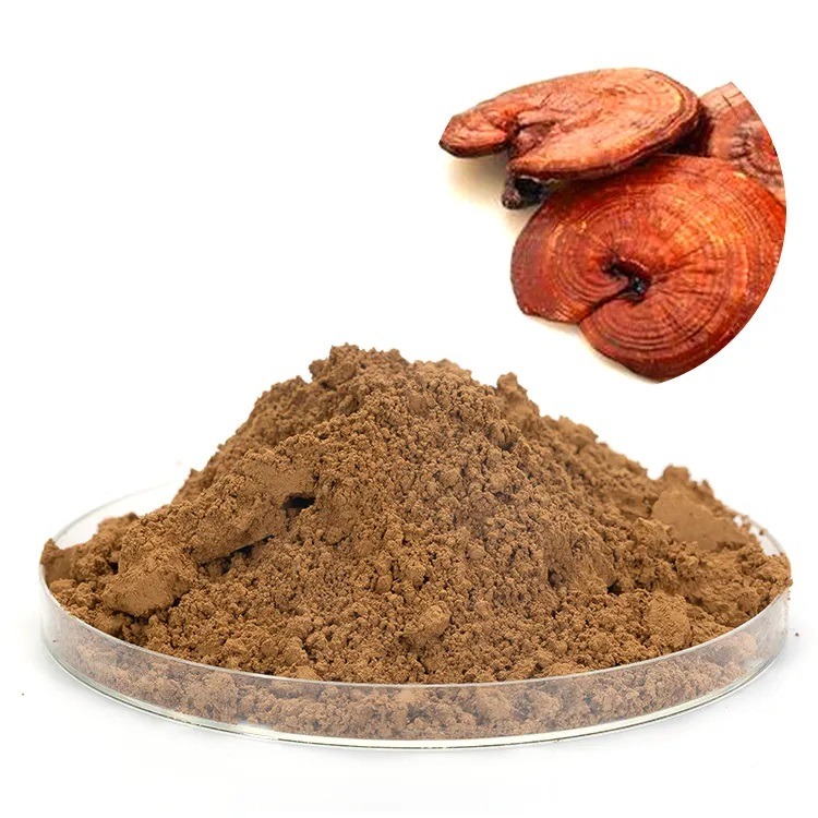
Our study showed the discriminating effect of reishi(ganoderma lucidum) extract against cancer cell viability. Reishi extract(1% cracked spores, 13.5% polysaccharides, and 6% triterpenes) can prevent cancer cell viability in 24 h. This compound’s studies have demonstrated similar effects on the MDA-MB-231 breast and PC-3 prostate cancer cell lines. This effect was not observed in a mammary epithelial cell line. Reishi only reduced cell viability by 11% at the highest concentration tested (1.0 mg/mL), suggesting that reishi selectively inhibits cancer cell viability.
Our data also shows that reishi extract induces apoptosis as demonstrated by annexin V and 6-CFDA staining paralleled with reduced expression of BCL-2, BCL-XL, and survivin in IBC(inflammatory breast cancer) cells after 24 h of reishi. Similar to these findings, many reports demonstrate that reishi causes cell cycle arrest at different stages and apoptosis and autophagy in cancer cell lines.
Our data showing a reishi-induced high decrease in cell viability coupled with increased apoptosis demonstrates that this mushroom extract may exert a stronger inhibitory effect on SUM-149 IBC cells than in other cancers. Most studies on the results of the reishi compound on cancer cell death attribute to apoptosis induction due to mitochondrial dysfunction caused by inhibition of key mitochondrial antiapoptotic proteins and increases in the BAX/Bcl-2 or BAX/BAX/Bcl-XL ratios.
A recent review reported that targeting cell proliferation pathways is the most promising directed therapy for IBC. Accordingly, our data show that reishi extract reduces the expression of Bcl-2, Bcl-XL, and survivin, which are key proteins for cancer cell survival.
Tumor invasion and progression are multifaceted processes that involve cell adhesion and proteolytic degradation of tissue barriers. IBC cells are thought to invade by passive metastasis, where cells secrete differentiation factors, stimulate vasculogenesis, and invade as a cluster of tumor cells (IBC spheroids), pathologically termed emboli, located within a de novo formed vessel. The embolus maintains cell-cell attachments as it moves through the vessel and lodges within a dermal lymph node, causing the inflammatory phenotype that is characteristic of IBC.
Reishi extract effectively inhibits the invasion of IBC cells in 3-D culture. Although the effects of reduced invasion may be due to reduced cell numbers due to cell death, the capacity of IBC cells to create the IBC spheroids was impaired by reishi extract. Since IBC tumor cell emboli are more efficient at forming metastases and are more resistant to chemo- and radiotherapy than single cells, it seems feasible to prevent IBC with a compound with anti-invasive properties that can disintegrate the cell spheroids.
IBC patient tissue biopsies overexpress E-cadherin, fibroblast growth factor (FGF2), and eIF4G. Because increased E-cadherin expression in IBC cells is correlated with cell adhesions that are required for invasion, we investigated the effect of reishi on the expression of this IBC biomarker. We show that reishi affects E-cadherin expression posttranscriptionally because reishi reduced E-cadherin protein expression without affecting CDH1 mRNA expression. Loss of E-cadherin in noninflammatory breast cancer results in EMT; this can increase cell motility, thus increasing invasion. However, in the unique phenotype of IBC, overexpression of E-cadherin to mediate the tight spheroids is necessary for the attack. Accordingly, our results show that inhibition of E-cadherin by reishi did not increase IBC cell invasion.
Moreover, downregulation of E-cadherin expression may result in nuclear accumulation of beta-catenin, leading to the subsequent activation of the beta-catenin/TCF (T cell factor) transcription complex, which are downstream components of the Wnt signaling pathway. We show slightly reduced beta-catenin protein expression and no nuclear localization upon reishi treatment. Moreover, the massive downregulation of gene and protein synthesis, and the accompanying apoptosis induction by reishi, strongly suggest that the beta-catenin-regulated pro-proliferative transcriptional activities are suppressed by reishi treatment.
Interestingly, reishi extract also inhibited the expression of the translation initiation factor, eIF4G. Recent studies demonstrate that eIF4GI silencing in SUM-149 cells reduces E-cadherin and p120-catenin protein (but not mRNA) expression and reduced invasion. Furthermore, in this study, they showed that overexpression of eIF4GI in IBC promotes internal ribosome entry site (IRES)-dependent translation initiation. eIF4GI increased mRNA translation was shown to be partly responsible for the unusual pathological properties of IBC: overexpression of E-cadherin, strong homotypic IBC cell interaction, formation of tumor emboli, and pronounced IBC cell invasion.
We demonstrate that reishi extract inhibits eIF4G, E-cadherin, and p120-catenin protein expression, which, combined with reduced cell viability, may account for the tumor spheroid disintegration, thus reducing cancer cell invasion.
 Reishi extract dramatically reduced the expression of genes involved in cancer cell survival, invasion, and metastasis. Reishi downregulated the expression of FGFR2 and PDGFB, which are genes involved in mitogenic signaling, and TERT, a gene involved in cell senescence. Studies on urothelial cells show that reishi induces apoptosis and inhibits telomerase activity, decreasing bladder cancer cell growth.
Reishi reduced the expression of CDKN2A, which is a cell cycle kinase inhibitor. Since cyclin-dependent kinases (CDK) are activated by various mechanisms, including phosphorylation and dephosphorylation events, decreased gene expression of CDK inhibitors may not necessarily result in CDK activation. Reishi upregulated expression of the IL-8 gene in IBC cells.
However, a study using the same extract on MCF-7 cells exposed to oxidative stress showed that Reishi reduced IL-8 secretion. Therefore, it is possible that although gene expression is increased, posttranslational processing, thus activity of this chemokine, is modulated by reishi in IBC cells.
Studies using a human inflammatory carcinoma xenograft (MARY-X), where homophilic tumor emboli were present within lymphovascular places, lead to reasoning that cell adhesion, angiogenic factors, and proteolytic enzymes released by tumor cells might facilitate intravasation.
Reishi extract downregulated the expression of MMP9 and inhibited MMP2 and MMP9 activities of IBC cells. These gelatinases are involved in proteolytic degradation of the extracellular matrix during tumor invasion. Studies using the same SUM-149 cell line show that similar to reishi extract, expression of a dominant-negative E-cadherin decreased IBC cell invasion via inhibition of ERK1/2 phosphorylation and decreased MMP-9 gene expression and activity.
Others have shown that reishi triterpene acid B inhibited MMP-9 expression, ERK1/2 phosphorylation, and subsequent suppression of activator protein (AP)-1 and NF-kB DNA binding activities.
Based on our findings, we conclude that reishi is a potent anti-invasion agent that prevents tumor spheroid formation with the potential to inhibit the spread of IBC. This action can be correlated with reduced viability and inhibition of eIF4G, E-cadherin, MMP-9, and p120-catenin, key proteins responsible for tumor growth and invasion in IBC. The selection of reishi extract was due to its current use by local naturopathic physicians in cancer patients. It has been shown to improve the quality, prolong patients’ lives, and not interfere with chemotherapy.
Therefore, our findings suggest that reishi extract could be used as a novel anticancer therapeutic for IBC patients.

Leave A Comment